
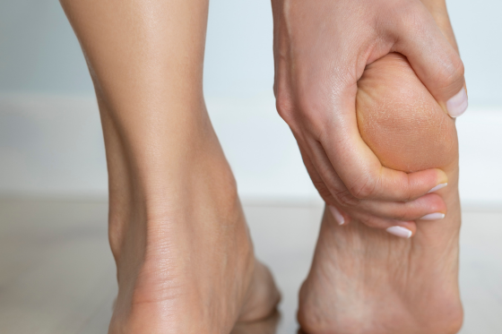
There’s a specific way this goes wrong.
Someone develops pain at the back of the heel. They look it up, find the standard Achilles exercise — heel drops off the edge of a step, letting the heel sink down below the level — and start doing them diligently. Three sets, twice a day, for weeks.
It gets worse.
They assume they need to push harder, or that they’ve been unlucky. What’s actually happened is that they’ve been doing an exercise designed for a different problem, and the specific thing that makes it useful for that problem is the thing making theirs worse.
Back-of-heel pain reverses several of the rules that apply elsewhere in the foot. Here’s why.
Two Achilles Problems, Not One
The Achilles tendon can develop problems in two distinct locations, and they’re considered separate clinical entities rather than variations on a theme.
Midportion tendinopathy occurs in the body of the tendon, a few centimeters above the heel bone. You can usually pinch the sore spot between your fingers, and the tendon may feel thickened there.
Insertional tendinopathy occurs within roughly two centimeters of where the tendon attaches to the heel bone — right at the back of the heel itself.
They can feel similar to the person experiencing them. They respond quite differently to treatment.
Alongside insertional tendinopathy, two other structures often join in: the retrocalcaneal bursa, a small fluid-filled sac sitting between the tendon and the heel bone, and in some people a bony prominence at the back of the heel — a Haglund’s deformity, sometimes called a pump bump. When these occur together the combination is often called Haglund’s syndrome.
Why the Standard Exercise Backfires
Here’s the mechanism, and it’s worth understanding because it explains everything else.
When you pull your foot upward into dorsiflexion — as you do at the bottom of a heel drop off a step — the back of the heel bone rotates toward the Achilles tendon. That compresses the retrocalcaneal bursa and the deep fibers of the tendon against bone.
For midportion problems, that’s irrelevant; the painful tissue sits well above the compression zone, and tensile loading through full range is genuinely useful.
For insertional problems, the painful tissue is precisely where the compression happens. Every repetition squeezes irritated tissue against bone.
The evidence reflects this. Systematic reviews of the standard eccentric heel-drop protocol have reported satisfactory outcomes in only around 28 to 32 percent of people with insertional problems — far below its performance in midportion cases. When researchers modified the protocol to eliminate dorsiflexion, using floor-level exercises only, reported satisfaction rose to around 67 percent.
Same exercise, one variable removed, roughly double the success rate.
The Rules That Reverse
Under the heel: load through full range. Back of the heel: limit the range.
Plantar fascia loading commonly uses a towel under the toes precisely to maximize tension. Insertional Achilles loading typically avoids taking the ankle past neutral into dorsiflexion. The general principle — progressive loading builds tolerance — still holds. The range in which you apply it flips.
Under the heel: stretching the calf often helps. Back of the heel: aggressive calf stretching often aggravates.
Limited ankle dorsiflexion is a recognized contributor to plantar heel pain, and calf flexibility work usually belongs in the plan. But the position that stretches the calf is the same position that compresses the Achilles insertion. Deep, forceful stretching with the heel down is one of the more common self-inflicted aggravators here.
Under the heel: firm, flat support usually helps. Back of the heel: a heel lift usually helps.
Raising the heel slightly reduces the amount of dorsiflexion the ankle travels through during normal walking, which reduces compression at the insertion. Heel lifts are a standard part of conservative management for this condition — and they’re the opposite of what you’d reach for with plantar heel pain.
Under the heel: barefoot on hard floors is the aggravator. Back of the heel: the back of your shoe may be.
A firm heel counter — genuinely useful for plantar problems — presses directly on the painful area in insertional cases, particularly where a bony prominence is present. Rigid dress shoes, ski boots, cleats, and stiff work boots are frequent culprits. Softer backs, or temporarily modified footwear, often bring meaningful relief.
Under the heel: it’s often an overuse problem in active people. Back of the heel: it’s frequently seen in less active people too.
Insertional presentations are notably common in people who aren’t training hard, which surprises those who assume tendon problems belong to athletes.
What Back-of-Heel Pain Feels Like
Typical features worth reporting accurately:
Pain right at the back of the heel rather than underneath. Stiffness and pain on the first steps in the morning and after sitting. Tenderness when pressing either side of the tendon just above the heel bone, or on the bony prominence itself. Pain that worsens going uphill, on stairs, or in certain shoes. Sometimes visible swelling, redness, or a firm bump at the back of the heel.
If your fingers can pinch a tender, thickened section of tendon a few centimeters above the heel, that points more toward a midportion problem — a different plan.
What Tends to Help
Load it, but within a limited range. Progressive calf and Achilles strengthening performed on flat ground, with the heel not dropping below level. Published approaches use relatively high repetition volumes across several months, progressing load as symptoms allow.
Use the next-day rule. Some discomfort during exercise is acceptable provided it settles by the following day. Symptoms clearly worse the next morning mean the load was too much.
Modify footwear. A small heel lift, and avoiding rigid heel counters pressing on the sore area while it settles.
Reduce the specific aggravators. Hill walking, stairs, and deep calf stretching, temporarily.
Be patient with the timeline. Conservative management for these presentations is generally trialed over a long period — often more than a year before surgical options are seriously considered. That’s slower than most people expect, and it’s the normal course rather than a sign of failure.
What to Be Careful About
Corticosteroid injection directly into the tendon is generally avoided in Achilles problems because of concerns about tendon degeneration and rupture risk. Injection into an isolated bursa is a different proposition and a discussion for a physician.
Sudden increases in load on a tendon that’s already irritated.
Pushing through sharp pain rather than working within tolerable discomfort.
When to Get It Assessed Urgently
Seek same-day care if you felt a sudden pop or a sensation of being kicked in the back of the leg, if you can’t push off or rise onto your toes on that leg, or if there’s a visible gap in the tendon. These can indicate an Achilles rupture, and prompt assessment matters.
Get assessed promptly for back-of-heel pain with redness, warmth, and swelling alongside fever; pain that’s severe and unrelenting at rest; back-of-heel pain in a younger adult with prolonged morning stiffness, back pain, other joint involvement, or psoriasis — inflammatory conditions commonly affect sites where tendons attach to bone, and the heel is among the most frequent; or heel pain in an active child or adolescent, which is usually a different condition entirely and needs its own assessment.
The Practical Takeaway
If you have back-of-heel pain and you’ve been doing heel drops off a step, stop and get it looked at before continuing.
The exercise isn’t wrong. It’s just built for a problem two inches further up the tendon — and in this location, the detail that makes it effective is the detail causing the trouble.
Get the Location Right First
Under the heel and behind the heel are different problems with, in several respects, opposite plans. Treating one as the other is a common reason people stall.
Highland Physical Therapy offers a free discovery visit at no cost and no obligation. You’ll get a proper examination that establishes exactly where the problem sits, an explanation of why your symptoms behave the way they do, and a loading plan built for your specific presentation.
If your presentation needs imaging or a physician’s opinion, we’ll tell you plainly.
